RESEARCH TEAMS
SKIN IMMUNITY AND INFLAMMATION
Our team brings together experts in immunology, inflammation and dermatology to investigate inflammatory skin diseases and the regulatory/effector adaptive immunity governing their pathophysiology. We explore key peripheral and skin mechanisms that regulate these diseases through state-of-the art cutting-edge technologies. Integrating “basic” and “translational” researchers in close collaboration with dermatologists and plastic surgeons, our team ultimately aims at identifying new disease activity biomarkers and at developing innovative therapeutic strategies.
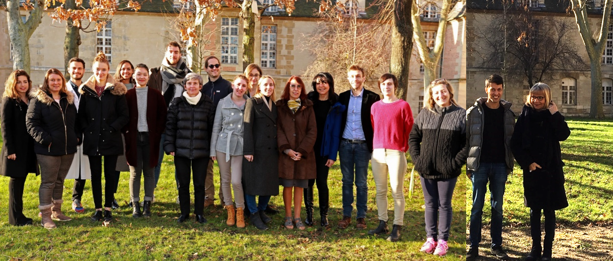
The team “Skin Immunity and Inflammation” aims at identifying the immunological pathways involved in the regulation of inflammatory skin diseases in the blood and in situ in the skin. Ultimately, we aim to pave the way for developing novel therapeutic strategies for skin diseases including auto- and alloimmune disorders with cutaneous manifestations (lupus, dermatomyositis, scleroderma, graft versus host diseases), inflammatory skin disorders (atopic dermatitis, psoriasis, neutrophilic dermatoses), cutaneous immunosenescence, and rare diseases of the epidermal barrier (hereditary epidermolysis bullosa). Our research is focused on regulatory T cells, regulatory B cells, skin fibroblasts, stem cells and their extracellular vesicles/exosomes and use in-vitro, ex-vivo and in-vivo experimental model systems combining cellular and molecular technologies including multicolor and mass cytometry, bulk and single cell RNA sequencing and multiproteomics. To reach its ultimate goal, this team brings together basic and translational researchers specialized in the skin, stem cells and immune cells (Drs Reem Al-Daccak, Hélène Le Buanec, and Laurence Michel), and medical clinical researchers being dermatologists (Prs Jean-David Bouaziz, and Vincent Descamps) or plastic surgeons (Prs Maurice Mimoun, Marc Chaouat, and David Boccara).

PROJECTS
Regulatory T cell populations in healthy and inflammatory skin diseases patients
Project managers: Hélène Le Buanec, Jean-David Bouaziz
The regulatory arm of the immune system is not only physiologically vital for the control of auto-reactive cells (Self-tolerance) and the modulation and ending of adaptive immune reactions (ergotypic immune reaction) but also pathophysiologically active during chronic inflammatory diseases including cancer, autoimmune diseases, viral infection and transplant organ rejection. The aim of this project is to define the phenotypically-distinct and functionally distinct T regulatory subsets in inflammatory skin pathologies. The ultimate goal is to produce and expand regulatory cell populations with stable regulatory properties under various inflammatory conditions.
Molecular Signature of skin chronic graft versus host disease
Project managers: Jean-David Bouaziz, Hélène Le Buanec, Gérard Socié
Graft-versus-host disease (GVHD) is a severe complication and a major cause of non-relapse mortality following allogenic hematopoietic stem-cell transplantation (allo-HSCT).
Cutaneous involvement of chronic GVHD has a wide range of manifestations including a lichenoid form currently defined by a mixed Th1/Th17 signature, and a sclerotic form defined by a Th1 signature. Despite substantial heterogeneity of innate and adaptive immune cells recruited to the skin and the different clinical manifestations, treatment depends mainly on the severity of the skin involvement, and relies on systemic, high-dose glucocorticoids alone or in combination with a calcineurin inhibitor. The aim of this project is to define and compare the transcriptome of lichen planus cGVHD and morphea cGVHD using RNA-Seq, in order to define common and unique inflammatory pathways in cGVHD that are both pathogenic and targetable
Role type 1 interferon subtypes in Dermatomyositis
Project managers: Jean-David Bouaziz, Hélène Le Buanec
Interface dermatitis of the skin is observed in auto-immune diseases such as lupus and dermatomyositis (DM) which may involve internal organs and skin injuries sensitive to UV rays. The pathophysiology of DM is poorly understood while it begins to be better described in lupus. Type I interferon pathway (IFN I) seems critical in the pathophysiology of these diseases and a new drug targeting type I interferon receptor has recently been proved effective for lupus treatment.
IFN-k is a subtype of IFN I produced by keratinocytes. Our research team has recently shown that IFN-k may have a key role in MDA5+ amyopathic DM (aDM), a severe clinical form of DM frequently associated with life-threatening pulmonary injury.
Type III IFNs (IFN-L) use the same signaling pathway than IFN I but their receptor is preferentially located in the skin and IFN-L could also be important players in ID mechanisms.
The objectives of this work are to clarify the role of type I and III IFNs in ID, especially in aDM and to study the effect of their inhibition in a mouse model of ID.
Mechanical and Molecular regulation of fibroblast and immune cell interactions in healthy and inflammatory fibrotic skin diseases
Project managers: Jean-David Bouaziz, Laurence Michel
Key words: inflammatory skin diseases, dermal fibroblasts, fibrosis, Keloids, cutaneous T lymphocytes
Inflammation is a common dominator of skin diseases and skin ageing (inflamm-aging). In the aged skin, dermatological diseases are associated with the presence and accumulation of senescent cells. Although cellular senescence is thought as a safeguard against malignancy development, accumulation of senescent cells in the skin is detrimental for neighboring immune and non-immune cells (Toutfaire, Biochem Pharmacol, 2017). Our data brought evidence indicating that fibroblasts from aged skin display altered senescent-like features, with reduced proliferative potential, inflammatory mediator production and senescent-associated secretory phenotype (SASP) factors that maintain an inflammatory chronic status within the tissue (Brun, Exp Dermatol, 2016). To reach precise definition of fibroblast contribution to skin ageing and associated diseases, as well as in fibrotic conditions, we aim to analyze:
- the whole secretome and proteome of dermal fibroblasts as well as their spatial and Single-Cell transcriptional profiling to identify functionally distinct human dermal fibroblast subpopulations in pathological diseases.
- the actin cytoskeleton, since the organization of the cell cytoskeleton and the extracellular matrix structure work in coordination and are major features in skin aging and fibrosis. This enables to determine whether skin aging alteration brings a decrease in fibroblast communication and cell motility together with their tissue migration within the environment. Traction Force Microscopy (TFM) will enable visualization of the dynamic characteristics of mechanical forces exhibited by young and old fibroblasts or fibrotic cells.
These approaches are currently extended to skin fibrotic diseases involving fibroblasts, such as scleroderma, graft-versus host disease cutaneous manifestations as well hypertrophic scars, with a focus on keloids. Co-cultures and 3-D bio-construct in vitro models are developed by combination of dermal fibroblasts and immune cells for immune interaction studies and further application to dermatological diseases. Skin biopsies from patients are currently used for cell isolation and (single-cell) RNAseq transcriptomic and proteomic analysis. Result validations are obtained using appropriate mouse models.
Current pharmacological studies derived from our approach of the interactions between the cutaneous microenvironment and the immune system, include:
- the in vitro study of the effects of calcium alginate wound dressings on the involvement of the innate immune system (NK cells, Macrophages M1/M2) in cutaneous wound healing process, with a focus on fibroblast stimulation;
- the proof of concept of the immunomodulating and anti-inflammatory activity of new Pickering Emulsions in vitro and in a preclinical study of imiquimod-induced psoriasis-like skin inflammation in mice in vivo.
Placental Stem cells and/or their released Extracellular Vesicles to mitigate Dystrophic Epidermolysis Bullosa symptoms
Project managers: Reem Al-Daccak, Jean-David Bouaziz
Epidermolysis Bullosa (EB) is a group of inherited skin diseases characterized by defective epithelial cell adhesion leading to skin fragility and trauma-induced blistering. Dystrophic EB (DEB), autosomal dominant (DDEB) or autosomal recessive (RDEB), is a major EB subtype. DEB patients skin displays an intrinsic proinflammatory state and therefore, DEB is currently viewed as a systemic inflammatory disorder. Because a real cure is still lacking, symptom relief therapies through promoting physiological healing of DEB are a current focus of research. This would involve a mutual control of the overlapping phases regulating proliferation and migration of cells needed for extracellular matrix (ECM) deposition and remodeling, angiogenesis, but also DEB inherent immune/inflammatory nature and injury-associated inflammation. In this context, stem cells as symptom relief therapy hold particular promise given their capacity to regulate/manage the diverse nature of DEB. Founded on our documented studies and steered by our long-standing know-how our project is devoted to provide the basic and translation knowledge that will support the development of placental stem cells-based strategy to overcome the devastating clinical symptoms of DEB disorder and to set the basis that would enhance the likelihood of their success in entering clinical investigation. Our working hypothesis is that “The interplay between placental stem cells and components of immune system constitute the backbone of healing, repair, and regeneration of injured DEB tissues”, which we challenge through 3 specific objectives:
- Define the immunological/inflammatory landscape of DEB patients.
- Modulate and polarize the intrinsic activated immune/proinflammatory landscape of DEB patients by allogeneic placental stem cells and/or their extracellular vesicles towards anti-inflammatory immune response of physiological wound healing and repair/regeneration.
- Highlight the mechanism-of-action: Metabolic reprogramming.
Immune regulation of atopic dermatitis and prurigo nodularis
Project managers: Reem Al-Daccak, Jean-David Bouaziz
Prurigo nodularis (PN) is a chronic skin condition of unknown etiology with an unmet therapeutic need. Indeed, the lack of approved therapies and the limited efficacy of off-label treatments reflect the current poor understanding of PN pathogenesis. The mechanisms triggering PN. Barrier dysfunction and T cell immune activation are the 2 main pathophysiological components in atopic dermatitis. The same is suggested for PN that may be the result of a neuro-immunologic dysfunction involving Th2 pathways. In this study we therefore, aim at exploring whether these entities share indeed a common pathogenesis by revealing the role of the Th2 pathway in PN pathogenesis. Our study might also pave the way for developing novel Th2-targeted therapeutic strategies for PN.

Principal Investigators
- Jean-David Bouaziz, MD, PhD, team leader, APHP/Paris University
Tél. : 01 42 49 43 91 - Reem Al-Daccak, researcher, INSERM
Tél. : 01 53 72 20 63 - Hélène Le Buanec, researcher, CNRS
Tél. : 01 53 72 20 80 - Laurence Michel, researcher, INSERM
Tél. : 01 53 72 20 55 / 06 75 04 62 03 - Vincent Descamps, Dermatologist, MD, PhD, APHP/ Paris University
- Maurice Mimoun, Plastic Surgeon, MD, PhD, APHP/ Paris University
- Marc Chaouat, Plastic Surgeon, MD, PhD, APHP/ Paris University
- David Boccara, Plastic Surgeon, MD, PhD, APHP/ Paris University


